Monday, February 13, 2012

Me, I, Oh Sis!

Wow dat title ^ I'm hilarious. Honestly like why am I so funny? People ask me all the time...


Okay, so this article talks about variation that occurs before and after mitosis and meiotic recombination. First, what is meiotic recombination? Meiotic recombination is one of the defining events in the formation of eggs and sperm. It is a process in which DNA is exchanged between "partner" chromosomes. If the process is distrubed, chromosomes often go astray during meiotic  division, resulting in eggs or sperm with too many or too few chromosomes. In humans, the resulting embryos are almost always abnormal and are a major source of miscarriages or congenital birth defects, such as Down syndrome.


This image displays the two methods used in meiotic recombination that I have described below.


Meiotic recombination, crossovers (exchanges of genetic material between homologous chromosomes. One of the final phases of genetic recombination, which occurs during prophase I of meiosis in a process called synapsis), and gene conversions (process by which DNA sequence information is transferred from one DNA helix (remainging unchanged) to another DNA helix, whose sequence is altered; one way a gene may be mutated) all affect variation and are therefore important from an evolutionary standpoint. Crossovers increase genetic diversity by redistributing existing variation and gene converstions alter the frequency of alleles. 
A group of researches conducted a series of experiments in which they sequenced Arabidopsis Landsberg erecta (Ler) and two sets of all four meiotic products from a Columbia (Col)/Ler hybrid to try and find the genome-wide variation. This would also help them find the meiotic recombination at the nucleotide resolution. 
Their results showed many single nucleotide polymorphisms (SNPs) which are  DNA sequence variations that occur when a single nucleotide in the genome differs between members of a biological species or paired chromosomes in an individual. They also found many different sized insertions and deletions, which matched the other SNPs throughout the genome. Using a mutant, they discovered that two sets of four meiotic products were produced. They were then analyzed by sequencing in a nonfungal species. 
A total of eighteen crossovers and four gene conversions revealed that Arabidopsis gene conversions are probably fewer, and have shorter tracts, than those in yeast.
In conclusion, meiotic recombination and chromosome assortment dramatically redistributed genome variations in cells that undergo meiosis. This plays a hand in population diversity! It presents a quick way to generate copy-number variation of sequences whose chromosomes are positioned differently in both Col and Ler.

Thursday, February 9, 2012

ProtoANKURgenes?

When Dwebs was going over oncogenes and protooncogenes in class, I was getting pretty confused. Thankfully this video on youtube helped me understand the difference between the two. It pretty much states that point mutations, one of the mutations we learned about, is one of the ways a protooncogene becomes an oncogene. The reason some cells become cancer cells is actually because protooncogenes become amplified. This pushes the cell towards uncontrolled growth. Another way cells become cancerous is when protooncogenes are rearranged through chromosomal translocation. This is when a gene from one chromosome is stuck to the promoter region of another chromosome. I would definitely recommend you watch this video if you were having trouble grasping the concept of protooncogenes and oncogenes.


In other news... this came up while I was on YouTube and I was like "aww, how relevant!" Not quite sure if I like it, perhaps it'll grow on me?

I Wish I Was a Dwarf!

This lengthy article is about a defective growth gene in people with rare dwarfism disorder that impedes their ability to get cancer and diabetes. This is truly miraculous considering how cancer and diabetes are two of the most common diseases that plague mankind.
Over the past few years, Jaime Guevara-Aguirre, has served as a physician in a small town in Ecuador where his patients stand at a mere 3'11". His patients have a rare genetic disorder known as Laron syndrome. A third of the world's population of people that have Laron syndrome reside in this remote village in Ecuador. What's so special about these cuties? Well, almost none of them suffer from cancer or diabetes!


This is a picture of Guevara-Aguirre (right) standing with one of his Laron syndrome patients.

These midgets have an error in their growth hormone receptor (GHR) gene which gives them their short stature. However, it also seems to keep them immune to diabetes and cancer! People who have this deficiency in growth hormone receptors are also unresponsive to growth hormone and have low levels of insulin-like growth factor 1 (IGF1). This is a hormone that promotes cell growth and inhibits programmed cell death.
Researchers conducted a series of experiments to investigate cellular response to IGF1. The studies set a precedent seeing as it was the first time that the GHR-deficiency mutation was being studied in humans. Because it is such a rare disorder, it was hard to study the subjects before. Results have shown that IGF1 can be regulated by diet aka IGF1 is an important determinant of cancer. 
In conclusion, mutations aren't always bad. Sure, people who have Laron syndrome are super short but hey, they will probably live longer and healthier lives than you will! Ain't that depressing :/

Oh, P53!

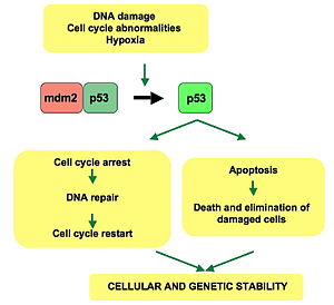
In chapter 14, we talked about tumor-supressor genes. A tumor-supressor gene is a gene that, under normal conditions, encodes a protein that prevents cancer. However, when a mutation eliminates its function, cancer may occur aka cancer-causing mutations in tumor-suppressor genes are due to a loss of activity. One tumor-supressor gene that we have talked about is p53. p53 is a transcription factor that acts as a sensor of DNA damage. It promotes DNA repair, prevents the progression through the cell cycle, and promotes apoptosis. It is present in the G1 phase of the cell cycle and controls the first checkpoint. About 50% of all human cancers are associated with mutations in this gene. This includes malignant tumors of the lung, breast, esophagus, liver, bladder, and brain, as well as leukemias and lymphomas.


This is a simple diagram that shows the pathway of p53.


This article talks about p53 and its molecular basis to chemoresistance in breast cancer. As you may have known, TP53 is the gene that encodes the tumor protein p53. Mutations in this gene have been known to be linked with resistance to anthracyclines (class of drugs used in cancer chemotherapy) and mitomycin (anticancer drug that belongs to the family of drugs called antitumor antibiotics) in breast cancer. This article goes over the possible responsibilities of different parts in the p53 cascade giving respect to drug resistance. The full article goes over the research that took place. It also talks about p53 activation in response to genotoxic stress and phsphorylations by ataxia telangiectasia mutated/ataxia telangiectasia and radiation resistance gene 3 related (ATM/ATR). Chk1 and 2 are also considered to be very important. A little while back researchers discovered that nonsense mutations in CHEK2 that encoded the chk2 protein, were found to predict resistance to anthracycline therapy in some tumors that contained wild-type TP53. As of right now, there is no evidence that MDM2 amplifications in breast cancers are resistant to anthracyclines. The roles of p53 isoforms and p53-induced transcription of non-coding RNA are yet to be determined. Experts say that disturbances affecting the p53 pathways may play key roles in chemoresistance in cancer. Although TP53 is not an exact marker for drug resistance, it still may be considered a signal for identifying critical gene cascades.

Monday, January 30, 2012

Mr. Anderson to the Rescue!

This is a video in which Mr. Anderson goes over gene regulation. He explains from the beginning, mentioning how E. coli is in our intestines and breaks down what we eat. He goes over and defines regulatory genes, regulatory sequences, promoters, lac operon, trp operon, transcription factors, and activators and repressors. He then carries out scenarios through diagrams so we can see how things like repressors and activators work. If you are having trouble grasping this concept, I highly recommend you watch this video!

Circadian Clock Without DNA

This article talks about two recent articles that were released recently. It goes into depth about both of the articles that have been causing quite the controversy. In both papers, both of the researchers' laboratory models were eukaryotic cells and neither had any DNA transcription or RNA translation going on inside the cell.
The results of both papers demonstrated that transcription of DNA and translation of RNA is not needed for the generation of circadian rhythms in two different types of eukaryotic cells belonging to evolutionarily very distant relatives – protists and mammals.
In the case of red blood cells, the result is lucid – there is no DNA or RNA in these cells. Thus, leaving the circadian rhythms in these cells, to be generated in the cytoplasm.
In the case of O.tauri, the picture result was a little bit more complex: the cells had a nucleus which had DNA. There was a clock driven by transcription and translation of recognized "clock genes." However, when this mechanism was supressed, by constant darkness or by chemicals, the cells still exhibited circadian rhythms generated by the molecules residing in the cytoplasm (and some of those molecules may have been strands of RNA transcribed earlier).
This is exactly what they discovered in both cases – there was a clear circadian rhythm of peroxiredoxins state-switching both in cultured red blood cells and in the cultured Ostreococcus tauri .
In sum, the phase at which the DNA-centered clock starts its cycle is determined by the phase of the cytoplasmic clock, not the other way round, i.e., the cytoplasmic clock is dominant over the nuclear clock.

Regulation of Transcription in Eukaryotes

This article talks about regulating transcription in eukaryotes. Controlling gene expression is far more complex in eukaryotes than it is in bacteria; but, they have the same basics. Eukaryotic gene expression is controlled at initiation of transcription. In bacteria, proteins control transcription. They bind to specific regulatory sequences and modulate the activity of RNA polymerase. The same thing happens in eukaryotes. The complex task of regulating gene expression in the many differentiated cell types of multicellular organisms is accomplished primarily by the synergy of multiple different transcriptional regulatory proteins. Packaging DNA into chromatin and modifying it by the process of methylation, transmit further levels of complexity to the control of eukaryotic gene expression.
This is a picture of a transcriptional activator. It has two independent domains.
This is a picture of a eukaryotic repressor. Some repressors block the binding of activators to regulatory sequences. While, others have active repression domains that inhibit transcription by interacting with general transcription factors. 


Gene expression in eukaryotes is regulated by transcriptional activators and repressors. Activators bind to regulatory DNA sequences and stimulate transcription. They appear to be modular proteins. So, the DNA binds and activates domains of different factors and thereby these factors can frequently be interchanged using different DNA techniques. Repressors on the other hand, bind to specific DNA sequences. I like to think of them as inhibitors. In most cases, eukaryotic repressors interfere with the binding of other transcription factors to DNA. For example, when a repressor binds near the transcription start site, it can block the interaction of RNA polymerase (or general transcription factors) with the promoter. Other repressors compete with activators for binding to specific regulatory sequences.