Wednesday, November 30, 2011

Understanding DNA Replication Control

This article talks about understanding how DNA replication is controlled. Completion of genome duplication during the S-phase of the cell cycle is crucial for the maintenance of genomic cohesion. The S-phase is where DNA replication takes place in the cell cycle. The cell also forms a second centrosome during this phase. The synthesis phase occurs after the first growth (G1) phase, and therefore about midway through interphase. At the start of the S phase, each chromosome has only one DNA molecule, but by the end of the S phase each has two, which, barring copying errors, are genetically identical, i.e. they have identical base sequences.


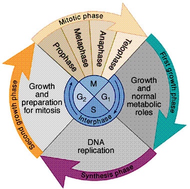
This is a picture of the cell cycle. In purple is the synthesis phase where DNA is replicated.


In eukaryotes, chromosomal DNA replication is accomplished by the activity of multiple origins of DNA replication throughout the genome. Origin specification, selection and activity, and the availability of replication factors and the regulation of DNA replication licensing have unique and frequent features that can be found amongst eukaryotes. Although the studies on the semiconservative nature of chromosome duplication were carried out in the mid 1950s in Vicia faba; plant DNA replication studies have been scarce. They have received a drive in the last decade, after the completion of sequencing the Arabidopsis thaliana genome, that hasn't been seen before, and more recently of other plant genomes. This past year, for example, has seen major advances with the use of genomic approaches to study chromosomal replication timing, DNA replication origins and licensing control mechanisms. In this minireview article is a discussion of the recent discoveries in plants in the context of what is known at the genomic level in other eukaryotes. These studies make up the basis for addressing, in the future, key questions about replication origin specification and function that will be of great importance for plants and for the rest of multicellular organisms.


Monday, November 14, 2011

A Different Perspective: Apoptosis

A fun little skit on apoptosis on the cellular level. 


This video starts off with giving various reasons why a cell might die. It then goes further into explaining what steps the cell takes, what enzymes bind where, and ultimately how the cell is destroyed. It also explains how the cell death affects it's environment. Overall, I found this to be very helpful and easy to understand.

Where and Why: Apoptosis

This article addresses two main questions:
Why are cells that die by programmed cell death generated?

Obviously, the answer would be different depending on different cell types but for the most part some cells are generated in excess and only those that become properly functional survive (as happens in parts of the nervous system). This is kind of like survival of the fittest because only the competent cells will survive. In some cases, the mechanism that generates cells that are needed also generates unneeded ones as well (as happens in the immune system). And some cells that die may be needed, but only temporarily.
 Cells die because they are harmful or because it takes less energy to destroy them than to keep them alive and healthy. As of now, programmed cell death has been known to occur only in animals, although it remains possible that bacteria, fungi and plants may also use similar processes to eliminate unwanted cells.
Why do these cells die instead of surviving?
One of the main reasons for cell death is to get rid of dangerous cells, the one's whose existence can harm the organism. Cells literally "kill themselves for the greater good". They could be mutants that can lead to cancer--apoptosis is therefore very important in the formation (or nonformation) of cancer. Also, positive and negative selection occur among the cells of the immune system. Cells that recognize "self" (ones that would attack the organism's own cells) are instructed to die during this process. Also, cells that are infected by a virus, can sometimes recognize the infection and kill themselves as to not allow the virus to be spread further.

Cell Apoptosis

Pulmonary emphysema is a chronic lung condition in which the alveoli are destroyed, narrowed, collapsed, stretched, or over-inflated. Over-inflation of the air-sacs is a result of a breakdown of the walls of the alveoli, and causes a decrease in respiratory function and breathlessness. Damage to the air sacs is irreversible and results in permanent "holes" in the tissues of the lower lungs.

This picture shows the difference between normal alveoli and alveoli with pulmonary emphysema.


This article talks apoptosis and it's role in pulmonary emphysema. Pulmonary emphysema is a powerful phenomenon that involves the gradual destruction of extracellular matrix by presence of an extra amount of proteases, but also apoptosiscellproliferation, and senescence (process of deterioration with age). Cellular proliferation makes up for enhanced alveolar cell death, whereas cell aging caused by cigarette smoking and increased cell turnover slows/stops cell proliferation, leading to apoptosis. As a result, alveolar cells gradually disappear and emphysematous lesions advance. At the same time, cellular senescence causes long-term inflammation through enhanced production of proinflammatory cytokines. Recent research suggests that DNA damage (double strand breaks) underlies the molecular mechanisms of these factors in the gradual destruction of extracellular matrix; apoptosiscell senescence, and chronic inflammation.

Wednesday, November 2, 2011

Cell Respiration Song


This is actually a very helpful video. It's based on a really catchy Black Eyed Peas song and has already helped me remember the steps of cell respiration. You'll have to watch/listen to understand. The song explains the 3 steps of cell respiration. It explains what the products of each step are. It also goes over the energy intermediates. It explains aerobic and anaerobic respiration. Overall, I thought this was a very clever and helpful video.

Lactic Acid ≠ Soreness

After a long day benching at the gym, I sometimes wonder why my arms are so sore... this article explains why. This article talks about why lactic acid builds up in muscles and why it causes soreness. As we start to perform more strenuous exercises, we breathe faster to attain more oxygen to keep our muscles working. Although the body prefers to generate energy using oxygen, sometimes our bodies require energy production faster than we can adequately deliver oxygen. Our bodies have to produce energy without the presence of oxygen. Through glycolysis, glucose is broken down or metabolized into a substance called pyruvate through a series of steps. When oxygen is limited, the body temporarily converts pyruvate into a substance called lactate, which allows glucose breakdown--and thus energy production--to continue. Sometimes the lactate levels get high because we cannot get enough oxygen over an extended period of time. This increases the acidity of the muscle cells. The same metabolic pathways that permit the breakdown of glucose to energy perform poorly in this acidic environment. This is a natural defense mechanism from the body. Eventually the body slows down, oxygen becomes available and lactate reverts back to pyruvate, allowing continued aerobic metabolism and energy for the body to recover.

 This is a diagram showing how lactate forms.
Lactic acid is not the cause of the soreness in the days to follow. Researchers who have examined lactate levels right after exercise found little correlation with the level of muscle soreness felt a few days later. Though the exact cause of delayed-onset muscle soreness is still unknown, most research suggests actual muscle cell damage and an elevated release of various metabolites into the tissue surrounding the muscle cells. These responses to extreme exercise result in an inflammatory-repair response, and are probably the  leading cause of the swelling and soreness that peaks a day or two after the event and resolves a few days later, depending on the severity of the damage.

Reduced Respiration Rates in the Periwinkle Littorina littorea

This article talks about how exposure to elevated temperatures and Pco(2) (partial pressure of carbon dioxide; is a blood gas test that checks for the amount of carbon dioxide gas that is in the blood) reduces the respiration rate and energy status in the periwinkle Littorina littorea. Scientists investigated the effects of elevated Pco(2) and temperature on the whole-organism and cellular physiology of the periwinkle Littorina littorea.
 
 This is a picture of  the periwinkle Littorina littorea

They chose to study this organism because in the future marine organisms will have to cope with multiple environmental changes associated with increased levels of atmospheric Pco(2), such as ocean warming and acidification. They wanted to find out how organisms will adapt to these changes and if they will survive. To understand this they would need an in-depth understanding of the physiological and biochemical mechanisms that are the basis of  organismal responses to climate change. Respiration rates, adenylate energy nucleotide concentrations and indexes, and end-product metabolite concentrations were measured. The final results were compared to the controls'. We saw that the snails decreased their respiration rate by 31% in response to elevated Pco(2) and by 15% in response to a combination of increased Pco(2) and temperature. We saw that the decreased respiration rates were associated with metabolic reduction and there was an increase in end-product metabolites in acidified treatments, indicating an increased reliance on anaerobic metabolism. They concluded that marine organisms will probably adapt in complex ways to future environmental drivers, which will likely have negative effects on growth, population dynamics, and, ultimately, ecosystem processes.